
The cannula is about a millimetre wide. The fiber inside it is thinner still — a strand of silica, sheathed, running from a console on the trolley to a tip you can never see once it is under the skin. On the console is a number: 10 W.
That number is identical whether the fiber ends in a plane-polished bare tip or in a ten-millimetre diffusing cylinder; identical whether the core is 200 µm or 1000 µm; identical whether the tip was fired clean thirty seconds ago or has spent four minutes carbonising against blood and liquefied fat. The console does not know any of this. It reports what it is sending.
Then the light arrives at the fat, and the number is not the same at all.
This article is about the last optical element in the chain — the fiber — and about treating it as the part of the system that sets the thermal field rather than as the tube that happens to carry the laser to the tissue. The argument is narrow and technical: at a fixed wavelength and wattage, the tip geometry decides the shape of the heat, the core diameter decides its density, and the condition of the end face decides whether either of those things is still true at minute four. Everything a lipolysis fiber datasheet lists — core, numerical aperture, tip form, jacket, connector — is one of those three.
A caveat before the physics, because this is an aesthetic procedure and the honest version matters. The published evidence does not establish that laser-assisted lipolysis (LAL) is superior to conventional suction-assisted fat removal, and one recent review found no clear skin-tightening benefit from endolaser either (see the section near the end). What follows is about how the laser delivers its energy into fat, not a claim that it delivers a better result than not using a laser. If a clinic runs a 980 nm or 1470 nm platform, the fiber is the largest single variable it still controls. That is the whole scope of this piece.
The cannula contains one fiber, and the fiber is the procedure
Strip the marketing away and an interstitial lipolysis setup has three elements: a laser console, a cannula that defines the tunnel and protects the tissue from the shaft, and a fiber threaded through that cannula. The console offers two or three wavelengths and a wattage. The cannula offers a diameter and a length. The fiber offers everything else — how the beam leaves, how concentrated it is, how it behaves against fat versus water, and whether it survives the case.
There is a reason this article puts the fiber at the centre rather than the wavelength. The installed base of aesthetic platforms is overwhelmingly built around 980 nm diode and 1470 nm diode, and the physics of that choice is settled: 980 nm is absorbed by both haemoglobin and water, so it coagulates small vessels while it heats fat, and 1470 nm sits on a much stronger water-absorption band, so its energy is more tightly confined near the tip (Mordon et al., Biomed Eng Online 2008; Wassmer et al., Photomed Laser Surg 2009). A clinic buys the console once. The fiber is what it buys every week. It is also the only component in the chain that touches the patient, that is selected per anatomy, and that can change its own behaviour halfway through a case.
The fine geometry is not incidental. The reference configuration in the modelling literature is a 1 mm cannula inserted into the hypodermis about 8 mm below the skin surface — that is what the finite-element models assume, and it is why the cannula length in a handpiece kit is a clinical decision and not a packaging one (Mordon et al., Biomed Eng Online 2008). Our lipolysis handpiece range runs cannula lengths from 100 mm to 300 mm, and a 2 m flexible version for facial work; the length sets how deep the fiber tip can reach before the hub hits skin, which is the difference between treating the fat you mapped and treating the fat you can reach.
Two lesion shapes from one console
The single most useful experiment on this topic is a 2013 bench study that fired a forward-firing fiber and a diffusing fiber into porcine fat and porcine liver at matched settings, and imaged the resulting lesions with a digital camera and a thermal camera (Kim, Park and Lee, Lasers Surg Med 2013).
The finding that matters is not a temperature — it is a shape, and the shape did not follow the beam.
| Forward-firing fiber | Diffusing fiber | |
| Emission geometry | one spot at the core face | a cylinder, 10–30 mm of emitting length |
| In fat | a reservoir of melted fat fed by a narrow channel: the beam tunnels in, the fat liquefies, and the ablation zone becomes roughly discoid, not a cone | a disc-shaped coagulated volume around the whole emitting length |
| In liver (water-rich) | an elongated ellipse — the classic “drilling effect”, a vaporised tunnel that advances as the dehydrated wall is ablated away | the same disc — tissue composition barely changed the outcome |
| Where the energy goes | concentrated at a face smaller than the core, so it reaches vaporisation temperature and the vapor cavity does the cutting | spread over a large area, so local intensity is far lower and the tissue is coagulated rather than vaporised |
Read the two rows about fat together. The same console, the same wattage, the same tissue: one fiber drills a channel and forms a molten reservoir at its centre, and the other lays down a fairly uniform coagulated disc with no channel at all. Changing the fiber changed the physical model of the procedure — from a cutting-and-melting tool to a volumetric heating tool. That is the sense in which the fiber sets the thermal field: it does not change how much energy is delivered, it changes the geometry of where it lands and how hot the interface gets.
Both ends of that product line exist for a reason, and they are not interchangeable: a diffuser fiber for a defined column of fat that should be heated evenly along its length, and a bare fiber for a forward, directed effect at a specific point. The one thing a forward-firing tip will never do is deposit energy evenly along its own axis, and the one thing a diffuser will never do is concentrate it at a point. Neither is a better fiber; they are two different geometries with two different thermal fields, and the console cannot tell them apart.
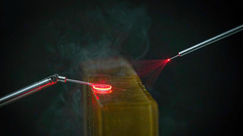
Core diameter: how a wattage becomes an irradiance
If the tip form sets the shape, the core diameter sets the density inside that shape — and this is the number most often missing from a case note, because it never appears on the console.
Irradiance is power divided by area, and area goes as the square of the radius. At a fixed 10 W, the same wattage produces these irradiances at the fiber face:
| Core diameter | Area at the face | Irradiance at 10 W |
| 200 µm | ≈ 0.031 mm² | ≈ 31,800 W/cm² |
| 400 µm | ≈ 0.126 mm² | ≈ 7,960 W/cm² |
| 600 µm | ≈ 0.283 mm² | ≈ 3,540 W/cm² |
| 1000 µm | ≈ 0.785 mm² | ≈ 1,270 W/cm² |
Two things follow, and both of them are counterintuitive enough to be worth stating as rules.
First: a bigger core is not a more powerful fiber — it is a cooler one, at the face, with a bigger hot volume. Going from 400 µm to 1000 µm drops the face irradiance by a factor of six at the same wattage. The extra energy is not lost; it is spread. A large core at moderate power is the tool for moving through a broad, soft area and heating a volume evenly. A small core at the same power is the tool for a fibrous, dense pocket where you need the tip to actually do something — and where, if you keep it moving, the higher irradiance is an advantage and, if you dwell, it is a burn.
Second: the fiber diameter you can pass is set by the cannula, so the two choices are linked. A 200 µm fiber inside a 2 mm cannula has room to sit where gravity and the tunnel put it; the same fiber loose in a much wider bore can bow away from the wall, and the tip is no longer where the depth readout says it is. This is the mechanical half of the “you cannot feel where the tip is” problem that every LAL review notes (McBean and Katz, J Clin Aesthet Dermatol 2011) — and it is a fit question, not a technique question.
Our lipolysis fiber range spans 200–1000 µm core in silica with standard FC and SMA 905 connectors, so the core is a per-case choice rather than a stock item, and the same handpiece can be run with the density you want for the tissue in front of it.