
Two operating theatres, same afternoon, same console model, same line in both op notes: 1470 nm, 8 W, 3 seconds. In one note the dose is written as ten shots per pile. In the other it is written as “until the cushion looked right.” Post-operative day one: VAS 2 and VAS 6. Nothing was different about the machine.
The console tells you four numbers — wavelength, power, pulse duration, and a running joule counter. It does not tell you how far the fibre has moved, how much of that energy landed above the dentate line, or how much heat the mucosa has already given back. Those three things decide the patient’s experience of the next two weeks, and none of them appear on any screen. This article is about how to reconstruct them, and about the two ceilings and one anatomical distance that the current literature actually supports.
If you want the fibre-level background first — why a radial tip lays down a cylinder rather than a point — that is covered in the radial vs conical tip comparison and in the general fibre tip design notes. This piece assumes that choice is made and asks the next question: how much energy, delivered where.
What a watt actually controls
Power is the rate of energy delivery. It is a measure of flow, not of reach, and the intuitive assumption — more watts, deeper burn — is the single most common misconception in LHP training.
An ex-vivo study using fresh porcine anorectal tissue puts a number on this. Fifty-four samples were randomised across 6, 8 and 10 W and 1, 2 or 3-second exposures, using a 1.85 mm fibre inserted the way it would be inserted into a pile. The longest lesion was produced by the longest exposure time — a mean of 3.93 mm at 3 seconds — with no significant difference between the three power levels (Danys et al., Open Medicine 2020). Depth tracked time. It did not track watts.
There is a second observation worth knowing because it inverts the expected direction. A morphological study at 1560 nm exposed haemorrhoidal tissue at 8, 9 and 10 W for 3 seconds and measured the damage: 8 W produced the deepest injury (up to 4 mm) with indistinct coagulation borders, 9 W produced 3 mm, and 10 W produced only 2 mm with sharp borders and minimal change in the surrounding tissue (Perm Medical Journal, 2026). At the highest power, the most pronounced reduction in node mass still occurred in the 8 W group.
The explanation is not exotic. At higher irradiance the tissue at the tip reaches vaporisation temperature faster; a charred, desiccated layer forms at the interface and begins absorbing and scattering the beam before it can travel. The energy is spent on the surface. You get a hotter tip, a smaller effect, and a black deposit on the glass — the classic precursor to fibre adhesion and burn-back.
Read both studies together and the practical rule falls out: watts set how fast you cook, seconds set how deep you cook. Power is the throttle. It is not the steering wheel and it is not the accelerator pedal.
Time, the pause, and the column you are actually building
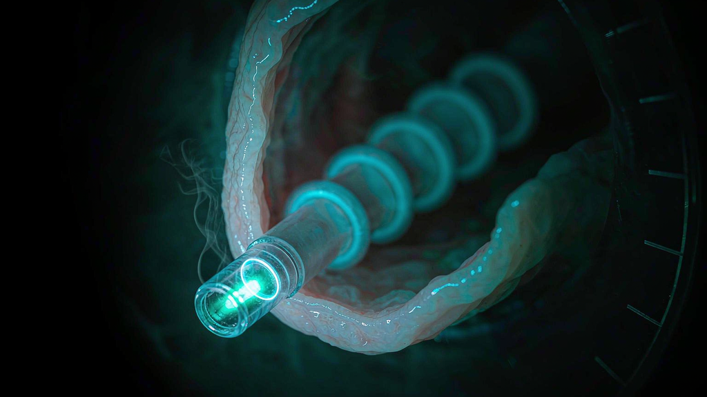
A radial tip does not drill a hole. It emits circumferentially, so as you withdraw it, it lays down a cylinder of coagulated tissue along the tract — and the length of that cylinder is set by how long the laser is on while the tip is stationary. This is also why the glass capsule on a purpose-built hemorrhoid fibre is dimensioned the way it is: a ~10 mm fused tip with a 1.5 mm outer diameter defines both the emitting length and the stiffness that lets the capsule track a submucosal tunnel without kinking.
That is exactly what Danys measured: a 3-second emission produced roughly a 4 mm column. The same authors’ recommendation for the next shot follows directly — begin the next application about 5 mm from the previous one. Five millimetres of spacing for four millimetres of lesion gives a chain of overlapping-but-not-stacked coagulation beads, which is what produces confluent shrinkage instead of a stack of isolated hotspots.
Now run the arithmetic on the classic protocol, because it is quietly self-consistent. Ten to twelve shots per pile, spaced 5 mm apart along the tract, treat roughly 5 to 6 cm of submucosal tunnel — the height of an average haemorrhoidal column plus a margin. At 8 W for 3 seconds, each shot is 24 joules, so a pile receives approximately 240 to 290 joules. Both the geometry and the dose land where the consensus says they should. Whoever wrote that protocol had already done this calculation.
The pause is the part that gets dropped in a busy list. Published descriptions alternate a 3-second emission with a 1-second interval, or use a duty cycle of roughly 2 seconds on and 1 second off. The purpose is not to rest the surgeon. At 1470 nm the optical penetration in water-bearing tissue is on the order of a couple of millimetres, so the subsurface tissue has absorbed nearly everything you sent in; the interval is the only mechanism giving that heat time to conduct away from the mucosa rather than accumulate at it. Remove the pause and you convert a deep coagulation into a superficial burn, with the same joule count on the counter — the machine will not notice, and the patient will.
Joules per pile: the number that does predict something
If there is one parameter that deserves to be written down in the op note, it is total energy per pile. The 2024 international LHP recommendations make the case explicitly: the number of shots per pile has never been defined — published practice ranges from 3 shots per pile to 10–12 — but “the total energy per pile may represent a better parameter as this has been somehow more uniformly reported across different studies,” and there is “a clear correlation between the amount of energy and the risk of morbidity.”
Those recommendations, developed by a group of LHP surgeons and voted on by 49 panellists with a Delphi process, set a ceiling:
Maximum energy per pile should not exceed 350 J. (strong consensus, 88.24%)
Note what that number does to the protocol above. The classic 10–12 shot sequence at 8 W × 3 s produces 240–290 J per pile. The consensus ceiling is not a new constraint invented by a panel — it is a formal confirmation of what the standard technique was already doing. That is worth sitting with, because it means the instinct to increase energy “to get a better result” is not an optimisation. It is a departure from the only dosing envelope with survival data behind it.
The ceiling measured in a different unit
There is a second number in the literature, and it is in a different unit. In a prospective cohort of 497 patients treated with a 1470 nm diode at the Siegen centre, the authors analysed day-of-surgery pain against a set of intraoperative parameters and found significant differences for mucopexy, for three treated segments, and — the only parameter that retained significance on its own — an energy level above 500 J (Weyand et al., Zentralbl Chir 2019). The paper’s conclusion is unambiguous: “the energy applied should be reduced to a minimum.”
Five hundred joules. But per what? Read the methods: the energy variable is what was consumed per patient across a mean of 2.7 treated knots. So the same cohort that produced the per-pile ceiling of 350 J also produced a per-case signal at 500 J — well below what three piles treated to the per-pile cap would consume.
This is the trap. If you plan case by case in per-pile terms, three piles at the cap is 1,050 J, which is not what Weyand’s cohort was describing when it flagged 500 J as the threshold for more day-of-surgery pain. Plan the dose in three units — per shot, per pile, per case — and write all three on the note. It costs nothing and it is the only way to see a dosimetry error while the patient is still on the table.
And the counter-evidence, which is equally real
Honesty requires the other side, because the correlation is not clean:
- A randomised comparison by Naderan and colleagues found no significant correlation between total laser energy and adverse outcomes following LHP — cited and echoed in a retrospective cohort of LHP combined with wedge resection, which found no difference in total energy between patients with and without complications (median 1,209.5 J vs 1,163.0 J, p = 0.28) and concluded that, within the standard operational range, laser energy is unlikely to be the primary determinant of complications or recurrence.
- A 100-patient multicentre Brazilian series of grade III disease found that higher total energy was associated with more complications from day 14 onward — but described the clinical effect as modest.
- A large single-centre series from Hanoi found mean energy ≥ 350 J per pile significantly associated with post-operative defecatory dysfunction (OR 1.91, 95% CI 1.02–3.56, p = 0.043) — and simultaneously found that the same threshold was not associated with urinary retention, which tracked anaesthetic technique and age instead.
So what is the honest position? Joules are not the dose. They are the accounting of the dose. Total energy is a weak aggregate that averages together two very different things: energy delivered at the right place, and energy delivered at the wrong place. A high joule count in the submucosa above the dentate line is not the same event as the same joule count lying against the anoderm. That is why a 1,200 J median can coexist with a clean complication profile in one series and a 500 J threshold can carry a pain signal in another — and it is why the next section matters more than either ceiling.
Why 2 cm above the dentate line matters
Two centimetres above the anal verge sits the dentate line. It is the most consequential landmark in the anal canal, because everything about the tissue changes across it.
Above the line: columnar epithelium, arterial supply from the superior rectal artery, venous drainage to the portal system, lymphatics to the internal iliac nodes, and autonomic innervation — insensate to cutting and to heat. Below the line: squamous anoderm, supply from the inferior rectal artery, drainage to the systemic circulation, lymphatics to the groin, and somatic innervation via the inferior rectal nerve — exquisitely sensitive.
That single division is the entire reason LHP is described as painless while a haemorrhoidectomy is not. It is not the wavelength. It is not the fibre. The “painless” property is a location, and the location is the tissue above the dentate line. A 1470 nm laser fired into the anoderm hurts exactly as much as any other 1470 nm laser fired into the anoderm.
So the operative rule is a placement rule. The fibre is introduced through a 2–3 mm microincision at or near the mucocutaneous junction — commonly 1 to 1.5 cm from the anal verge — and tunnelled submucosally in a straight line until the tip lies at the apex of the column, 2 to 3 cm above the dentate line, where the feeding pedicle is. Energy is then delivered on withdrawal, working down the tract. Straight trajectory, because a curved tunnel either wanders into the internal sphincter or, at worst, through the rectal wall; the reported cases of fistula-in-ano and rectal perforation after LHP are attributed to probe trajectory rather than to dose.
Three things follow, and the third one is the part that gets missed.
First, the first shots should be the ones with the most clearance. They are the furthest above the line, in tissue with no somatic supply, treating the pedicle where the arterial inflow actually is. That is where the case’s therapeutic value sits — the same principle as haemorrhoidal artery ligation, which is why several published LHP protocols deliberately borrow the technique’s logic. Joules spent here buy shrinkage. Joules spent near the line buy pain.
Second, the anoderm is not a target. The external component and any skin tag live below the line, in somatic territory. Weyand’s group makes the point directly: energy should be minimised to avoid perianal oedema and thrombosis, especially in the external component. If your dose plan involves “finishing” the external part with the same settings you used above the line, you have found a reliable way to produce the two complications that a laser procedure is supposed to avoid.
Third — and this is the non-obvious one — 2 cm is a window, not a direction. The 2024 recommendations do not simply say “stay high.” Their standard LHP sequence places 2 pulses about 0.5–1 cm above the dentate line, 3 pulses at the level of the dentate line, and 3 pulses below the dentate line, then permits extra pulses according to pile size and experience. Eight pulses at 24 J is about 190 J — comfortably under the per-pile ceiling with room to add.
Note that the ceiling and the sub-line pulses are doing two different jobs. The sub-line shots are the ones with the least margin for error and the least therapeutic return, so they should be the shortest emissions on the withdrawal, not the longest. And there is an upper edge too: the anal transition zone immediately above the dentate line carries the sensory mucosa that contributes to fine continence — sampling and discriminating between gas, liquid and solid. You do not want to be generous with energy in the transition zone either. The target window is the column above it, where the pedicle is. Treat the apex, spare the transition zone, touch the anoderm as little as the technique allows.