
Two fibers arrive in the same sterile pack. Both are 600 µm. Both carry an SMA 905 connector. Both are labelled for laser hemorrhoidoplasty at 1470 nm. The only thing that differs between them is about ten millimetres of glass at the distal end — and that ten millimetres decides whether the node in front of you shrinks evenly around its whole circumference, or whether you cook a single point on it while the rest of the cushion stays untouched.
Most LHP training material treats the fiber as a consumable you attach to the console. It is not. It is the last optical element of the laser system, and for a 1470 nm diode it is the only element that shapes where the energy lands. The generator gives you watts. The tip decides what those watts do.
This is the question we work through with proctologists who are specifying fibers for LHP — the same logic that sits behind the hemorrhoid fiber range we build and the more general fiber tip design principles that apply across every specialty we supply.
The only real difference is the distal geometry
Strip away the packaging and both fibers are the same object: a 600 µm (or 950 µm) pure silica core inside a cladding, terminating in roughly 10 mm of fused glass with an outer diameter of about 1.5 mm, transmitting above 90% at 980 and 1470 nm. The dimensions come from the anatomy, not from marketing — that glass capsule has to travel through a submucosal tunnel in the anal canal without kinking, and 1.5 mm is the practical compromise between stiffness and the size of the tunnel a proctoscope can give you.
What changes between the two products is how that glass capsule treats the light on the way out.
A radial tip converts the beam into a ring. The light leaves perpendicular to the fiber axis, all 360 degrees of it at once. Ten millimetres of glass becomes a ten-millimetre-long cylindrical emitter.
A conical tip refracts a large part of the beam sideways. This is where the brochures and the optical bench disagree, and it matters enough to spell out — we will come back to it in the next section.
Everything else in this comparison follows from those two sentences.
The radial tip: a ring, not a point
Put a plane-cleaved bare fiber in tissue at 3 W and the surface power density at the tip face can approach the order of 1 kW/cm². That is vaporisation territory. Tissue crosses from coagulation into boiling and charring faster than most operators expect, and once carbon forms on the tip face the tip becomes a broadband absorber — the fiber stops being an optical device and becomes a hot needle. That mechanism is documented in the venous literature and we have written about it at length in Radial vs Bare-Tip Fiber in EVLA, because it is the same physics wearing a different anatomical hat.
Spread the identical power over a circumference instead of a point, and the interface irradiance drops by orders of magnitude — into the regime where tissue coagulates steadily instead of exploding. The clinical consequence is not subtle.
The cleanest evidence we have for tip geometry as an independent variable comes from outside proctology, and it is worth importing. Ashpitel and colleagues took ten segments of human great saphenous vein and treated them at the same wavelength (1470 nm), the same power (10 W) and the same linear energy density, changing only the fiber — a forward-firing jacket-tipped fiber versus a radial-firing fiber. The radial fiber produced deeper thermal penetration and no carbonised tissue at all. The forward-firing fiber produced significant char. Same energy, same wavelength, same tissue. The only thing that moved was the delivery geometry.
That is the case for the radial tip in LHP, stated honestly. In a submucosal tunnel you cannot see, working within two or three millimetres of the anal mucosa, the difference between “coagulates evenly” and “chars a point” is the difference between a patient who goes back to work and a patient who comes back with an ulcer.
There are two things the radial tip cannot do, and both are worth knowing before you standardise on it:
- It cannot be aimed. It emits in every direction, including toward the mucosa. Position is everything, and you cannot compensate for a bad position by redirecting the beam — only by withdrawing and repositioning.
- It cannot treat a short segment. Ten millimetres of emitter treats ten millimetres of tissue. On a small node, that is longer than the structure you are trying to shrink.
Which brings us to the tip that most operators reach for in exactly those two situations.
The conical tip: what the geometry actually does
Search for a description of a conical hemorrhoid fiber and you will find the same phrase repeated across vendor pages: “focuses the beam forward for deeper penetration.” The measured polar diagrams are more interesting than that.
A group measuring eight multimode fibers with a 1064 nm diode recorded the emission profile of each tip geometry. Plane-polished fibers produced a single central lobe — a forward beam with an approximately Gaussian angular distribution, widening as numerical aperture rose. Conical tips produced two opposite lateral lobes with a minimal axial component. The cone geometry refracts the guided light sideways. It is not, optically speaking, a forward-firing tip at all.
So why do conical tips cut deeper than bare fibers? Because of the second mechanism, which has nothing to do with beam direction: the cone concentrates energy at a very small contact area where it meets tissue, and being in contact, it transfers heat efficiently by conduction. In porcine gingiva at 940 nm, conical tips cut 2,393 ± 468 µm deep compared with 711 ± 268 µm for a plane-cleaved bare fiber at the same 5 W, with more regular cuts and better handling — while the thermal damage zones around the cuts were comparable. Same laser, same power, roughly three times the cutting depth, purely from tip shape.
Put those two findings together and the conical tip’s clinical identity is clear. It is the tip that works tissue locally: it penetrates the submucosal tunnel more easily because of its tapered, sharp distal end, it can be directed at a specific structure rather than a volume, and it can divide or ablate rather than merely coagulate.
The canonical description of a conical-tip LHP technique comes from Filippou’s account of laser sclerotherapy of hemorrhoids, and one detail in it tells you everything about working with this geometry. The probe carries a bare fiber inside a special conical glass tip — wide illumination from the cone, sharp distal end for penetration. The laser runs at just 6 W, single 3-second shots. And during each shot, the probe is gently rotated around its own axis, explicitly to distribute the light evenly and prevent the fiber from adhering to the tissue. The tip is then moved 3 mm and the shot repeated, starting distal and withdrawing to the entry point, until the whole pile has been treated.
Read that rotation step twice. When you are using a radial fiber, you never think about adherence, because the light leaves in every direction and no single point takes the load. When you are using a conical tip in contact with tissue, adherence is the failure mode you are managing shot by shot. The technique exists to work around the geometry.
Note also the energy: 6 W. Experienced operators using a conical tip are routinely running at roughly half the wattage of a radial-fiber protocol, and reporting notably faster recovery and less perioperative pain as a result.
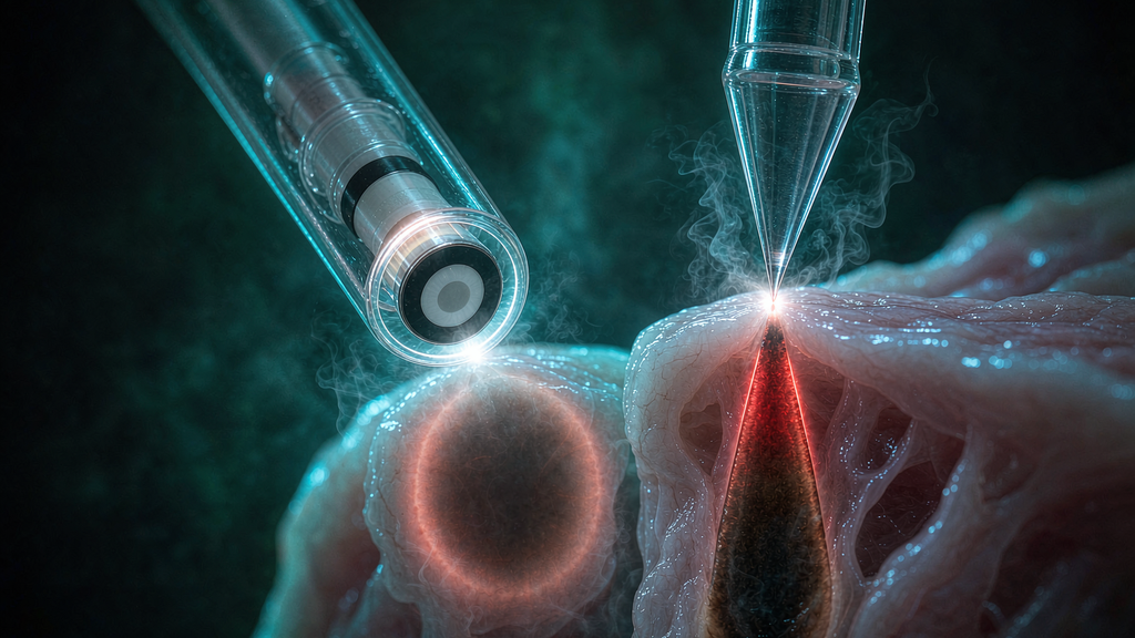
Side by side, on the variables that decide the case
| Radial tip | Conical tip | |
| Emission pattern | 360° ring, perpendicular to the fiber axis | Two lateral lobes with minimal axial component, plus high contact density at the cone |
| Coagulation zone shape | Cylindrical / annular, centred on the fiber | Focal, pear-shaped around the cone tip |
| Interface power density | Low — distributed over the whole circumference | High — concentrated at a small contact area |
| Directional control | None (cannot be aimed) | Yes (can be pointed at a structure) |
| Best target | Node volume reduction, whole-circumference shrinkage | Small nodes, the feeding pedicle at the base, skin tags and external components |
| Typical energy protocol | 8–12 W, 2–3 s pulses, 200–600 J per pile | 6 W, single 3 s shots, move 3 mm between shots, rotate during each shot |
| What it forgives | Being slightly off-axis in a blind tunnel | Being used on a small, localised target |
| How it fails | Under-treatment of the mucosal side; can’t be redirected | Carbonisation and adherence at the contact point; uneven dosing if you don’t move methodically |
| Manufacturing cost | Higher (fused cap, welded ring, polished emission window) | Lower (taper forming, single-point geometry) |
Which tip for which pile
This is the answer the title promises, and it separates into four situations rather than two.
Small node, Grade I–II, bleeding or discomfort but no prolapse → conical, low energy. You are working on a defined structure, not a volume. Six watts, three-second shots, 3 mm steps, rotate every shot. A radial fiber on a node this size treats more tissue than exists.
Medium to large node, Grade II–III, bleeding as the main complaint → radial. The objective here really is volumetric: shrink the cushion around its entire circumference while the mucosa above it stays intact. This is where the 360° ring earns its cost. Keep the withdrawal slow and deliberate, deliver the dose in a defined number of shots rather than “until it looks right,” and apply the ice finger afterwards to control edema.
Grade III with significant prolapse → radial plus mucopexy. The laser shrinks the cushion; it does not lift a prolapse. If the tissue needs to come back up, suture it back up — this is standard practice in the laser series that report acceptable recurrence rates.
Grade IV, irreducible prolapse, or a mixed internal/external component with a fibrotic skin tag → LHP is the wrong operation. We will say this plainly, because it is the single most important thing on this page: the published randomised data puts recurrence after LHP at 10% and, in a larger comparative series, at 21.6% versus 8.1% for Milligan-Morgan — and the recurrences cluster in the higher grades and in patients with significant prolapse. LHP is a superb operation with a bad reputation created by surgeons who used it in cases it was never designed for. Choose the right tip, and then choose the right patient.
A hybrid is legitimate, and more common than the literature admits. Many experienced operators use a conical tip to reach the apex and to coagulate the arterial supply at the pedicle, then switch to a radial fiber for bulk reduction of the cushion. There is no rule against two fibers in one case. If your console and your budget allow it, this is often the most controlled way to work a large node.
There is one more variable that interacts with tip choice and almost never gets discussed: the tip geometry changes how forgiving the blind tunnel is. You cannot see where the fiber is. A radial tip emits in every direction, so being a millimetre off-axis costs you almost nothing. A conical tip is directional, so being a millimetre off-axis means the dose lands where you did not intend — and the mucosa, which has no somatic pain fibres above the dentate line but plenty of sensitivity at the anoderm, will let you know in the recovery room. Match tip to your confidence in the position, not just to the node.