
Two weeks after a turbinate reduction, the patient is back in the chair and says the nose is blocked again. There is something in it. The endoscope shows what the discharge did not: a pale, shrunken turbinate with a grey-brown crust riding over the medial surface, thickest over the anterior third where the fiber spent the longest.
Nothing went wrong. The turbinate is smaller, which was the point. The crust is the reduction, still in progress — the dead layer the laser created is being carried off, and the nose is doing what a nose does with a necrotic surface.
The list of things blamed for that crust is long and mostly unhelpful: the patient did not irrigate, the power was too high, the air is dry, the technique was wrong. It usually misses the one thing that actually sets it. Crust is dried slough from the tissue you killed. Its thickness and its duration are decided by how much tissue that was, and how much of it was surface — and both of those are delivery decisions made partly at the console and partly at the tip of the fiber.
This article is about the three dials that set them — wavelength, delivery mode, and beam geometry — and about why “use less power” is not the answer most surgeons assume.
An honest caveat before the numbers: the title says “without the crusting”, and nothing in this specialty delivers that literally. You cannot shrink a turbinate by coagulation and have no slough. What you can do is keep the crust inside the one-to-two-week window most patients tolerate, instead of the six-week-plus window that a third of patients in some series still have at three months. That is a delivery question, and it has an evidence base.
The healing clock starts the moment the fiber fires
The histology of a diode-laser turbinate is unusually well described, and it explains the whole clinical course. In the Munich series that established 940 nm as the standard endonasal wavelength, 76 patients were treated in non-contact mode with a 940 nm diode at 10 W and 5 kJ per turbinate, and the treated turbinates were examined directly. What the laser produced was a horizontal ablation line 200–300 µm thick, bordered above and below by a coagulation zone — and that zone was the point of the procedure, because it is the tissue that later organises into the scar that shrinks the turbinate.
The clinical timetable followed the histology. The necrotic layer “detaches from the healthy underground” at about four weeks, and during the first two to four weeks nasal obstruction tracked the extent of postoperative oedema and nasal crusting — in other words, the patient’s symptom at week three was a read-out of the lesion created on day zero (Janda et al., Lasers Surg Med 2000). A later histological series in which 980 nm diode was applied submucosally under general anaesthesia found the same endpoint from the other direction: at three months the turbinate had become predominantly fibrous tissue, with the seromucinous glands and venous sinusoids that had made it bulky largely gone (Mohamed et al., Lasers Med Sci 2022).
Two consequences follow from that, and they are the reason this is a dosing article rather than a technique article.
First, crusting is not a complication in the usual sense; it is the visible stage of the mechanism. It is the slough of the layer you deliberately killed. Which means the honest question is not “how do I avoid crusting” but “how thick a layer am I willing to create, and for how long.”
Second, the reported healing windows across the literature differ by a factor of several, and they track the energy deposited rather than the wavelength name:
| Study / setting | Mode | Oedema | Crusting | Source |
| 940 nm diode, 10 W, 5 kJ/turbinate | non-contact | — | obstruction correlates with oedema + crust for 2–4 weeks; slough detaches ~4 weeks | Janda 2000 |
| 940 nm diode, 8–10 W | non-contact | — | 3–4 weeks | Sroka 2007 |
| 2100 nm Ho:YAG, 0.8–1.2 J, 4–8 Hz | non-contact | — | 1–2 weeks | Sroka 2007 |
| 940 nm diode, 42 patients | non-contact | resolves < 1 week | resolves by 6 weeks | Caffier 2008 |
| KTP 532 nm, 6 W CW, 400 µm fiber | — | — | 46.7% at 1 week → 16.7% at 1 month → 0% at 3 months | Vijayakumar 2016 |
| 940 nm diode, 60 patients | office, local | none observed | 19/60 (32%) still crusting at 3 months | Gupta 2019 |
Read the bottom row before the top ones. Three studies report crusting gone by four to six weeks; one reports a third of patients still crusting at three months. Same wavelength family, same indication, same “minimally invasive office procedure” — and a difference in outcome big enough that it changes what you tell the patient in the consent conversation. The difference lives in the dose, and the dose lives in the delivery.
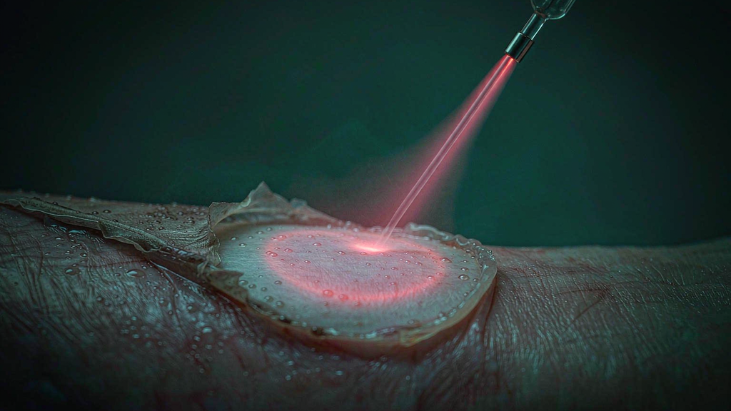
The clearest randomised evidence for less crusting is a wavelength change — and it is really an energy change
The strongest single piece of evidence on turbinate crusting is a trial design that removes almost every confounder: an intra-individual, prospective, randomised, double-blind comparison, in which each patient’s two nasal cavities were treated with two different wavelengths in the same sitting, by the same operator, in the same mode, through the same fiber core, with an independent physician grading the endoscopic photographs.
Twenty patients with hyperplastic inferior turbinates were treated endoscopically in non-contact mode — 1470 nm at 4–5 W on one side, 940 nm at 12 W on the other, both through 600 µm fibers. Follow-up was on days 1, 3, 7, 14 and 21 with rhinomanometry, the SNOT-20 questionnaire and photo documentation (Havel et al., Lasers Surg Med 2011).
The published result: operation time was significantly shorter on the 1470 nm side, and the volume reduction and symptom relief were equivalent. The group’s own paired dosimetry report makes the mechanism visible: 2.54 minutes and 125 J per turbinate at 1470 nm, against 3.30 minutes and 816 J at 940 nm (Havel et al., Medical Laser Application 2009). And the endoscopic assessment found that the 1470 nm side showed significantly less scab formation (p < 0.01) and better healing (p < 0.05) than the 940 nm side in the same nose (Havel et al., Lasers Surg Med 2014).
Sit with the numbers rather than the wavelength names. Both sides were non-contact, both used the same fiber core, and both reduced the turbinate to a comparable degree. The only things that differed were the wavelength and, as a consequence, the energy: one-sixth the joules for the same clinical result. The in-vitro arm of the same work explains why, and it is not a subtle effect — to produce the same macroscopically visible tissue effect, the 1470 nm system needed a reduced energy factor of 5–10× on perfused liver and 10–20× on muscle compared with 940 nm, because 1470 nm sits on a water absorption band that 940 nm does not.
So the correct reading of that trial is not “1470 nm is gentler.” It is “1470 nm can produce the same shrinkage with a fraction of the energy, and crusting tracks energy.” The lesion is thinner because there is less heat to dissipate, less tissue dies, and less slough has to come off. This is also why the wattage on the two sides differed by a factor of three and pointed the wrong way relative to crusting: the high-wattage side crusted more, because it also delivered six times the joules.
That is one clean lever. The counterweight is equally important, and it comes from the only placebo-controlled trial in turbinate surgery. Ninety-eight patients were randomised to diode laser, radiofrequency, microdebrider-assisted turbinoplasty, or a saline placebo injection, and followed with VAS scores and acoustic rhinometry. All three active techniques beat placebo — and, crucially for this article, nasal discharge and crusting decreased significantly in all four groups, including the placebo arm, with no difference between them (Harju et al., Laryngoscope 2018). At one year, that group found no deterioration in crusting, nasal discharge or mucociliary function in any arm (Am J Otolaryngol 2021).
Put the two trials side by side and you get the honest position, which is more useful than either alone. In the same nose, a wavelength-and-energy change measurably reduced scab formation. Across a whole population, a large share of the crusting after turbinate surgery is the natural history of an instrumented nose that is healing — the placebo arm crusted too, and then it stopped. Delivery choices modulate a process that is going to happen anyway. They matter most in the patients who crust worst: the ones with allergic mucosa, the ones in dry climates, the ones whose crusting is still there at three months.
Where the fiber enters: face irradiance, standoff and tip state
Everything above is decided at the console and by the wavelength — except that it is not, quite. The fiber decides how much of the console’s setting actually arrives, and in what shape.
The core diameter sets the irradiance at the face. Ten watts through a 400 µm core is about 7,960 W/cm²; through 600 µm it is about 3,540 W/cm². The console shows the same number in both cases. If a department standards on a small core for its fine work and then uses it for turbinate coagulation at turbinate wattages, it has quietly raised the surface fluence by more than a factor of two and pushed the tip toward carbonisation — which is a crust generator, and which the procedure does not need. The published turbinate protocols cluster on 400–600 µm for exactly this reason, across both the diode and the Ho:YAG literature.
Numerical aperture sets how forgiving the standoff is. Once the tip leaves the tissue the beam diverges, and density collapses with distance:
| Standoff (400 µm core, 10 W) | NA 0.22 | NA 0.37 |
| At the face | ≈ 7,960 W/cm² | ≈ 7,960 W/cm² |
| 2 mm | ≈ 750 W/cm² (9% of face) | ≈ 320 W/cm² (4% of face) |
| 5 mm | ≈ 180 W/cm² (2.3%) | ≈ 66 W/cm² (0.8%) |
The same geometry is described in the dental laser literature, where fluence falls by roughly 68% at 2 mm and 78% at 3 mm of tip-to-tissue distance. The physics is general: a millimetre of hand movement is worth more than a watt of console setting. This is the single most likely explanation for two operators in the same department getting different crusting profiles from the same protocol. A high-NA fiber spreads faster and is less sensitive to small changes in standoff, which makes it the more repeatable choice for non-contact turbinate work across multiple hands; a low-NA fiber keeps density concentrated, which is an advantage for a small precise target and a liability when the same hand is doing a whole turbinate.
Tip state is the third door, and it is the one that is easiest to overlook. A carbonised, initiated tip used in “non-contact” mode is not non-contact optics: the beam is now radiating from carbon, which is hotter and dirtier, and it will track a deeper and more irregular lesion than a clean face at the same setting. A chipped or freshly re-prepared face scatters, and scattered light produces focal hot spots — which is how you get an isolated crust and, at the other end, a synechia between the turbinate and the septum. The thirty-second aiming-beam check — a crisp, evenly bright, circular disc, with no rays, halo or double image — is therefore a crusting-control step as much as a safety step, and it belongs in the turbinate workflow for the same reason it belongs in the airway workflow.
Our bare fiber range covers 400 and 600 µm with both 0.22 and 0.37 NA and a plane-polished face in single-use and reusable versions; those two variables are the ones this section turns on, and buying them as one undifferentiated line item is how a department loses the ability to explain why one operator’s turbinates heal differently from another’s.