
A 9 mm great saphenous vein, fiber tip parked 2 cm below the saphenofemoral junction, tumescent already in, and two fibers on the trolley. One is a plane-polished bare fiber. The other has a fused quartz cap that throws light sideways. You pick one, you press the pedal, and for the next few minutes you are committed to a particular way of injuring a vein wall.
Most of the radial-versus-bare debate is conducted as if one fiber closes veins and the other does not. That is not the argument. Both close veins — reliably, and in published series at rates above 99%. What the tip geometry changes is how the wall is injured, and how the injury is distributed. That distinction turns out to matter a great deal for the patient’s week after the procedure, and much less than most people assume for the twelve-month duplex result.
This is the question we work through with clinicians specifying fibers for endovenous work — the same logic behind the radial fiber range and the bare fiber range we build. There is a fuller overview of how these fibers are matched to procedures on the vascular surgery page.
Two geometries, one photon budget
Start with the physics, because almost every clinical difference downstream traces back to it.
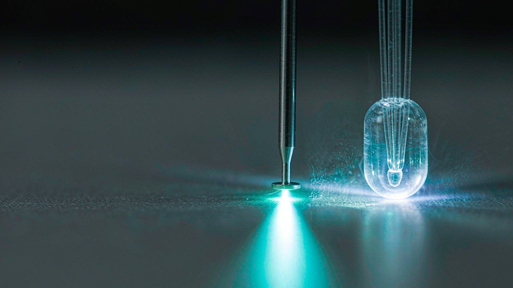
A bare fiber is a plane-polished silica face at the end of the fiber. Light leaves it forward, in a cone whose divergence is set by the numerical aperture — typically 0.22, giving roughly a 12–13° half-angle in air and a wider cone in blood and tissue. The energy density is highest right at the tip face and falls off with distance and with the square of the cone’s spread.
A radial fiber replaces that flat face with a fused quartz cap engineered so the light exits laterally around the full circumference — true 360° emission. On our radial fibers, the emitting window runs along a defined length of the distal glass (the reason the marked and unmarked variants exist), the fused ring is welded to the fiber body as a single mechanical unit, and transmission through the assembly stays above 90%.
Here is the sentence that gets lost: the photon budget is the same. Same wavelength, same watts, same joules. Both fibers put the same energy into the vein over the same pull-back distance at the same speed. The only variable that changed is where that energy lands first — a small forward-facing point, or a cylinder pressed against the wall.
From that single change, everything else follows.
What the wall actually sees
The mechanism of EVLA was worked out in bench and ex-vivo studies in the 2000s, and the numbers are worth knowing because they explain the clinical data later.
Disselhoff and colleagues, working with an 810 nm diode on ex-vivo human vein segments, measured intravascular temperature directly and found two things that reframed the field (Phlebology 2008;23(2):69–76):
- Tip temperatures reached 1200 °C. Not 100 °C, not 200 °C. A point-contact fiber in blood reaches temperatures at which tissue does not coagulate, it chars.
- The controlling mechanism is carbonization of the fiber tip, not steam. A carbon layer forms on the fiber face, and carbon is a broadband absorber — it converts the light into heat at the tip itself. The fiber stops behaving like an optical device and starts behaving like a hot needle. Their Schlieren imaging showed heat spread was tightly localised around the tip and dissipated almost immediately, which is exactly the behaviour you do not want from a treatment whose goal is a uniform circumferential wall injury.
A companion review by Fan and Rox-Anderson (Phlebology 2008;23(5):206–213) confirmed the histology: EVLA with a bare tip produces a transmural vein wall injury, typically with perforations and carbonization, and the injury is eccentric — maximal along the path of the laser contact. They also put a number on the steam theory that had been used to explain EVLA for years: steam production accounts for roughly 2% of the applied energy dose, and is therefore not the primary mechanism. Peak tip temperatures above 1000 °C with sustained temperatures of at least 300 °C in the firing zone are, and those are set by the contact point.
That is the bare-tip mode of action in one line: a high-temperature photothermolytic process at the point of contact between fiber and vein wall. It works — 1200 °C will close any vein it touches. But where it touches, it perforates; and where it does not touch, the wall may be under-treated. The eccentricity is built into the geometry.
A radial tip removes the point contact. Light is emitted through the quartz cap into the wall, and at 1470 or 1940 nm it is absorbed by the water in the wall tissue rather than by the blood in the lumen. Because the emission surface is a circumferential window rather than a flat face, a black carbon layer does not form on it in the same way — and the tip is not driven to 1200 °C by its own absorbed light.
One honest qualification, because the marketing version of this story overshoots: a radial fiber does not lower the delivered energy, it redistributes it. The thermal dose to the wall is comparable. What changes is the peak — from a point to a ring, from a needle to a cylinder.
The geometric trade-off the datasheet does not headline
There is a second difference between the two fiber families that gets far less attention than emission geometry, and in practice it decides more cases: outer diameter and stiffness.
| Bare fiber (600 µm typical) | Radial fiber | |
| Emitting surface | plane-polished face, forward | fused quartz cap, 360° |
| Typical outer diameter | 0.6–1.0 mm | 1.2–1.3 mm glass OD |
| Trackability in tortuous veins | better — flexible, slim | stiffer; harder past tight bends and aneurysmal segments |
| Access / introducer size | smaller sheath | needs a larger introducer |
| Contact behaviour | point contact; perforates on contact | rounded cap; resists perforation on contact |
| Tip cost | lower | higher |
A 600 µm bare fiber tracks through anatomy a 1.3 mm capped fiber will not. Tortuous below-knee segments, small saphenous veins, densely aneurysmal trunks, and the multiple-puncture workflows of a hybrid case are all places where the slim fiber wins on mechanics alone — and no amount of emission geometry compensates for a fiber that will not reach the segment you need to treat. The flip side is that once a bare tip is in place, every contact event is a potential perforation and a potential bruise.
The marked-ring variant of the radial fiber exists for the third dimension of this trade-off: depth control. Visible markings let you position and track the pull-back against the ultrasound image rather than by feel, which is how a defined retraction speed stops being a matter of opinion. If you are auditing why two operators using the same console get different bruising rates, this is usually where the difference lives.
The variables that decide more than the tip
Here is the part that gets buried under fiber marketing: in EVLA, the tip is not the largest lever you have. Four other variables move the endpoint more.
1. Wavelength. At 1470 and 1940 nm, the absorption target is water in the wall; at 810–980 nm, it is haemoglobin in the lumen. The water-specific wavelengths were introduced precisely because the haemoglobin-specific ones produced higher perforation and bruising rates. If you are comparing a 980 nm system with a 1470 nm system, you are comparing wavelengths, and the fiber is a detail.
2. LEED — and the fact that LEED alone is a meaningless number. Linear endovenous energy density is power divided by pull-back speed. Reported alone it tells you nothing: 50 J/cm is achieved by 10 W at 2 mm/s, or by 0.1 W at 0.02 mm/s, and the second case produces no thermal effect at all. Published protocols use nothing like that range, but the point stands — LEED must always be reported with the power and the speed that produced it. Practical published settings cluster around 8–12 W at 1470 nm with LEED in the 60–85 J/cm band; 1940 nm protocols have used 8 W with 160 J/cm over the first 5 cm and 80 J/cm thereafter. Whatever your protocol, it should be a written number, not “moderate pull-back”.
3. Tumescent infiltration. The perivenous fluid halo is what keeps heat away from skin and nerve. In the studies where it was introduced systematically, paraesthesia rates fell — from 15% to 9% in the radiofrequency series that first documented it, with a corresponding reduction in perivenous temperature. Tumescent volume and evenness belong in the same conversation as fiber choice.
4. Tip position relative to the junction. The convention of parking the tip 1.5–2 cm distal to the saphenofemoral junction exists to keep thermal energy out of the deep system. At 1470 nm the reported EHIT rate is around 2.4% versus 6.0% at 810 nm, and risk rises with vein diameter above about 7.5 mm, higher CEAP class, and simultaneous phlebectomy. That is a positioning and technique story, not a fiber-tip story — but it is a far bigger clinical event than a bruise.
So how should you actually choose
Default to the radial fiber for truncal work — GSV, SSV and anterior accessory saphenous vein ablation. Not because it closes veins better, but because it closes them with an order of magnitude less bruising and pain in the same-wavelength comparisons, and because the rounded cap is more forgiving when the vein moves. For a patient paying out of pocket and returning to work, that difference is the product. For large truncal diameters, where ESVS 2022 places endovenous thermal ablation at class IIa even above 12 mm, the centring effect is doing real work.
Choose the bare tip deliberately, not by default — where cost per case is the binding constraint, where the anatomy is small or tortuous enough that a 1.2–1.3 mm glass cap will not track, and where you are treating many short segments and tributaries rather than one long trunk. It is a legitimate, well-documented option: 355 limbs at LEED ≤80 J/cm with a 1470 nm bare tip produced 100% occlusion at one year. It is simply less forgiving of sloppy speed control and tip contact.
Never conclude anything from a mixed comparison. Same wavelength, same LEED, same pull-back speed, same tumescent protocol — then the fiber is the only variable left, and you can start attributing differences to it. Change two of those at once and you have a case series, not an answer.