
The console is set to 10 W. The duplex shows an 8 mm great saphenous vein running from groin to knee. Somewhere between the groin and the knee, the operator has to decide how fast to pull the fiber — and that decision, not the wattage on the screen, is what the vein wall actually feels. Two operators with the same laser, the same fiber and the same vein can deliver doses that differ by a factor of three, purely through pullback discipline.
Energy dosing is the least standardized number in endovenous laser ablation. Surveys of real practice report LEED anywhere from 30 to 120 J/cm for the same anatomy. This piece pulls the published numbers into one place, shows why the arithmetic behind them is simpler than the debates suggest, and separates the settings that follow from the fiber you are holding from the ones that follow from the anatomy in front of you.
Watts are a rate. J/cm is a dose. Pullback speed converts one into the other
The console displays watts because watts are what the diode produces. But ablation of a vein segment is determined by energy deposited per unit length — the linear endovenous energy density, LEED, expressed in J/cm. The three numbers are locked together by one identity that every EVLA operator is implicitly solving, whether or not they write it down:
Power (W) = LEED (J/cm) × pullback speed (cm/s)
This is not an analogy or an approximation — it is dimensional arithmetic. A 6 W laser pulled at 0.2 cm/s deposits 30 J/cm. The same 6 W pulled at 1 mm/s deposits 60 J/cm. Nothing changed on the console; the dose to the vein doubled.
Real protocols resolve this identity from both directions. The Samsung Medical Center group in Seoul ran a 1470 nm protocol at a fixed 6 W with a constant 2 mm/s pullback — which is simply 30 J/cm stated as a machine setting (Park et al., Surg Today 2015). The German four-zone study fixed the pullback at 1 mm/s and varied power by anatomical zone instead (Setia et al., J Clin Med 2023). Different habits, same equation.
It also means the endless argument about “how many watts” is incomplete on its own. Ten watts sounds aggressive; at a brisk 2 mm/s pullback it is a moderate 50 J/cm. Five watts sounds gentle; parked at 0.5 mm/s it is 100 J/cm and the patient will feel it. A wattage without a pullback speed is not a protocol. It is half of one.
What the dose range looks like across real practice
The published numbers are wide, and the width itself is informative. A national survey of Japanese institutions found mean LEED of 76.7 J/cm with a range of 50–120 J/cm at a mean power of 10 W — most of it delivered through 980 nm bare fibers, continuous mode, at the high-dose end of the spectrum (Japanese Vein Study XVII). Early high-LEED practice with 810–980 nm bare fibers routinely ran 80–120 J/cm.
Two things have been pulling the numbers down since. The first is wavelength: moving to 1470 nm, with its higher water absorption, let groups achieve occlusion at substantially lower doses — one randomized comparison of 1470 nm tumescence protocols still used 114–115 J/cm and reported 100% occlusion with less pain than the 810–980 nm literature, but later series settled far lower. The second is tip geometry: radial-emitting fibers spread the energy around the circumference instead of concentrating it at a carbonized tip, and the protocols that adopted them dropped accordingly.
| Configuration | Typical power | Typical LEED | Source |
| 980 nm, bare tip (early practice) | 10–14 W | 80–120 J/cm | Japanese Vein Study XVII; JPMA review |
| 1470 nm, bare tip, thigh GSV | 9–12 W | 80–100 J/cm | ESTLA technique, Phlebology 2023 |
| 1470 nm, radial, thigh GSV | 5–8 W | 50–70 J/cm | ESTLA technique, Phlebology 2023 |
| 1470 nm, radial, low LEED + high ligation | 6 W | 30 J/cm | Park et al., Surg Today 2015 |
| 1940 nm, radial, zone-dosed GSV | 2–4 W | 22–42 J/cm | Setia et al., J Clin Med 2023 |
Read the table as one trend rather than five protocols: every step that moves the energy off a carbonizing tip and into the vein wall — longer wavelength, radial emission — buys the same occlusion at a lower dose. The 1940 nm radial figures at the bottom of the table are not a different species of medicine; they are the same endpoint reached with less of the collateral heating that generates postoperative pain.
The fiber tip is part of the dose calculation
The reason a radial fiber runs 50–70 J/cm where a bare tip runs 80–100 J/cm is geometric. A bare fiber deposits energy forward through a carbonized tip that reaches roughly 1200 °C; the vein wall is heated indirectly, by conduction from that point source. A radial fiber emits 360° around the circumference, spreading the same joules over a cylinder of wall instead of a point, so the wall reaches ablative temperature without the tip paying for it in char.
For dosing, the practical consequences are specific:
- The same J/cm is not the same treatment. A bare tip concentrates its energy on a carbonized point; a radial fiber spreads it around the full circumference, cutting the fluence — energy per unit of wall area — to roughly one-fifth to one-ninth of the bare-tip value at equal power. That is the mechanical reason the radial protocols in the table above run 30–50 J/cm lower, and why protocol numbers only transfer between tip geometries with a deliberate adjustment.
- Low dose has a floor. The Samsung group paired their 30 J/cm protocol with surgical high ligation of the saphenofemoral junction precisely because low LEED alone leaves more partial-occlusion risk early on — 84.5% full occlusion at one month in their series, reaching 100% by six months (Park et al., 2015). Early attempts to run radial fibers without tumescent anesthesia, at a mean of just 22.7 J/cm, were abandoned after frequent recanalization (Japanese radial-fiber series).
- Tip construction matters at the same diameter. Burn-back behavior varies between fibers of identical nominal size, and a fiber that chars mid-pullback changes the effective dose the wall receives for the remainder of the segment. What to check on the datasheet is covered separately in the delivery fiber spec guide.
The morbidity side of this comparison is not theoretical. In a Japanese multicenter randomized trial comparing 1470 nm radial against 980 nm bare-tip fibers, postoperative bruising was recorded in 7.0% of the radial group versus 57.1% of the bare-tip group (Ann Vasc Dis, 2015) — and the earlier Doganci randomized trial found the same direction: thirteen patients with ecchymosis and fourteen with induration in the 980 nm bare-tip arm, against minimal findings in the 1470 nm radial arm, with better VCSS scores through the first month (Doganci & Demirkilic, EJVES 2010). Occlusion was not the differentiator in either trial — it was what the patient saw in the mirror on day five.
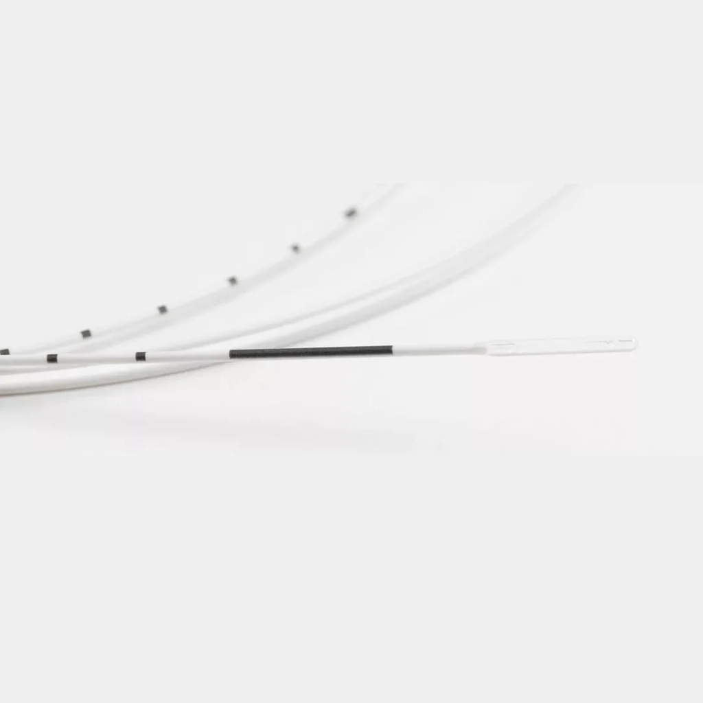
We make both geometries — the 360° radial fiber in 400/800 µm and 600/950 µm variants, ring-marked or unmarked, and the flat-polished bare fiber — and the dosing difference between them is exactly the 30–50 J/cm gap in the table above. The full physics of why is in our radial versus bare-tip comparison; this piece stays on the numbers.
Pullback speed: the dial nobody admits to setting
Ask what pullback speed a protocol uses and the honest answer in many departments is “whatever feels steady.” The equation says this is where the dose actually lives. The published protocols cluster at 1–2 mm/s, but the spread within that band is the entire dose:
- At 8 W: 2 mm/s gives 40 J/cm; 1 mm/s gives 80 J/cm. Same console, same fiber.
- The German zone-dosed study capped pullback at 1 mm/s and still only needed 2–4 W to hold 22–42 J/cm with 98.9% one-month occlusion (Setia et al., 2023).
- At the other extreme, an Indonesian crossectomy series pulled at 0.14 cm/s — one centimeter every seven seconds — at 40–60 J/cm, deliberately concentrating energy at the junction (single-center series, 2025).
There is also a thermal argument for the slower end that is independent of total dose. Heating is not instantaneous: the vein wall needs a finite residence time for conduction through the wall thickness and the surrounding tumescent halo. A high-wattage, fast-pullback protocol and a low-wattage, slow-pullback protocol can post identical LEED numbers while producing different axial temperature profiles — the fast pass under-treats the wall and over-treats the blood, part of why some groups report fewer side effects as watts came down even at constant dose.
Continuous or pulsed: the delivery mode changes the dose too
Most of the numbers above assume continuous emission, which is how the procedure is almost universally done now — but the pulsed protocols of the early EVLA era obey a different dose table. In the classic mathematical model of the procedure, permanent wall damage in a 3 mm vein required 65 J/cm in continuous mode but only 15 J/cm in pulsed mode; for a 5 mm vein, 100 versus 50 J/cm (Mordon et al., Biomed Eng Online 2006). Pulsed delivery stacks the energy into discrete shots, each of which dwells long enough to do more thermal work per joule — but it also demands that the fiber be repositioned precisely between pulses, and skipped segments and wall perforation under pulsed firing are the reasons continuous pullback displaced it.
The long-term data settle it the same way. A randomized trial comparing 12 W intermittent withdrawal against 14 W continuous pullback reported five-year outcomes favoring the continuous arm — better sustained occlusion and lower recurrence without added morbidity (Carradice et al., 2018). Whatever your wattage, the steady, boring, continuous pull is what the five-year duplex pictures were taken of.
One reporting convention deserves retirement here: LEED alone, without tip geometry and pullback speed, does not describe a treatment. Some groups add the endovenous fluence equivalent — energy normalized to the vein’s cross-sectional area — precisely because a fixed J/cm under-doses a 9 mm vein relative to a 5 mm one. The diameter adjustment below is the practical version of the same insight, and further down we put actual numbers on it.
The anatomy overrides the formula
The J/cm arithmetic assumes a uniform cylinder. Legs are not uniform, and the published dose modifications track three anatomical facts:
Vein diameter — and the EFE threshold underneath it
The German four-zone protocol adds 1 W for veins over 10 mm and subtracts 1 W when fiber sticking is encountered — a direct feedback control rather than a fixed table. The same logic appears across series as a rule of thumb: bigger wall, more watts, same speed.
The quantitative basis for that rule is the endovenous fluence equivalent, EFE — total energy divided by the inner vein wall surface, in J/cm². In the study that defined it, 263 great saphenous veins treated at 940 nm were split by dosing: the low-dose cohort averaged 12.8 J/cm², the high-dose cohort 35.1 J/cm², and their twelve-month occlusion curves separated permanently — 82.7% versus 97.0% (Proebstle et al., J Vasc Surg 2006). Durable occlusion tracked an EFE above roughly 20 J/cm².
That threshold converts directly into the LEED adjustments above. The inner surface of one centimeter of vein is π × diameter, so an 8 mm vein needs 20 × π × 0.8 ≈ 50 J/cm, and the rule of thumb that falls out is about 6.3 J/cm for every millimeter of vein diameter — 10 mm → 63 J/cm. The Japanese surveys reporting means near 77 J/cm were, in effect, dosing an average 11–12 mm vein past the threshold. The diameter adjustments in real protocols are this arithmetic done in the operator’s head.
Distance from skin
Near the knee and below, the vein sits closer to skin and saphenous nerve territory. The ESTLA protocol caps the lower leg at 40 J/cm regardless of what the thigh received, and doses tributaries purely by skin distance: 50–70 J/cm when the target is more than 10 mm deep, 20–40 J/cm when it is closer. Tumescent volume is the other lever here — the halo is doing thermal insulation work, not just anesthesia.
The junction
The last centimeter at the saphenofemoral junction is the most common failure point, and protocols handle it deliberately: the crossectomy series above gave a fixed 150 J bolus at the junction before starting the pullback, while others rely on flush tip placement at the junction instead. Either is a defensible convention; improvising it is not.
| Segment (GSV, radial 1470 nm) | Typical dose | Why |
| Thigh, upper (near junction) | 60–70 J/cm | Largest diameter, lowest recurrence stakes if under-treated |
| Thigh, mid and below-knee | 50–60 J/cm | Standard dose band |
| Lower leg | ≤40 J/cm | Skin and nerve proximity |
| Tributaries >10 mm deep | 50–70 J/cm | Depth-protected |
| Tributaries <10 mm deep | 20–40 J/cm | Resect or phlebectomy anything closer |
A worked example
Putting it together: an 8 mm GSV, thigh segment 35 cm, 1470 nm laser, 600/950 µm radial fiber, SMA 905.
- Target dose: 60 J/cm thigh average, capped at 40 J/cm below the knee.
- Chosen pullback: 1 mm/s — slow enough for wall conduction, fast enough for a reasonable case time.
- Power from the identity: 60 J/cm × 0.1 cm/s = 6 W. For the 8 mm diameter, that is consistent with the radial 1470 nm band (5–8 W); a vein over 10 mm would take 7 W instead.
- Below the knee, either drop to 4 W at the same speed, or keep 6 W and speed the pullback to 1.5 mm/s — the equation does not care which lever you move, but the monitor does: keep an eye on the steam envelope either way.
- Expected delivered energy: roughly 35 cm × 60 J/cm ≈ 2,100 J for the thigh, logged per segment.
- Sanity check against the EFE threshold: at 8 mm, 60 J/cm works out to about 24 J/cm² of wall — comfortably above the 20 J/cm² durability line. Had the vein measured 12 mm, the same 60 J/cm would be only 16 J/cm², below threshold — which is exactly why the diameter adjustment exists.
The point of writing the case out is that every number above is now a decision someone made before the first pulse — diameter adjustment, junction handling, dose cap by segment — instead of a wattage that felt right on the day.
What failure looks like in both directions
Under-dosing announces itself at follow-up: partial occlusion and early recanalization. The Samsung series is the cleanest illustration — their deliberately low 30 J/cm protocol needed the surgical ligation to cover the junction, and still showed 15.5% partial occlusion at one month before closing at six months. Under-dosing rarely causes a complication; it causes a second procedure.
Over-dosing announces itself in the recovery room. Numbness around the knee appeared in 31% of the Samsung patients at one month (resolving to 8.6% by six months), and laser-induced paresthesia in the German series ran 2.2% at one month with 0.5% persistent at a year — at zone doses under 42 J/cm. Endovenous heat-induced thrombosis, the extension of tail thrombus into the deep system, sat at 1.6%. Every one of these tracks dose and depth, which is why the interesting question about a protocol is never “what wattage” but “what dose, at what speed, at what depth from skin.”
The Brazilian randomized comparison is worth a final mention because it resists the neat narrative: 7 W at a mean 33.5 J/cm versus 15 W at 88.7 J/cm, and the high-dose group showed better stump stabilization and less reflux at six months (Araujo et al., J Vasc Bras 2016). With bare tips at older wavelengths, more energy did buy more secure closure — the lower doses in the table above are a dividend of tip geometry and wavelength, not a law of physics. If you are running a bare tip at 980 nm, the 50 J/cm mindset is not yet yours to have.
One further caveat belongs here, because it cuts against several rows of the comparison table at once. Pool the EVLA literature and closure sits around 92% regardless of wavelength, fiber or dose — the technique works almost whatever you do to it, which is precisely why sloppy comparisons are so easy to publish (Malskat et al., Lasers Med Sci 2014). The same review makes a point that deserves a wider audience: in most “longer wavelength hurts less” trials, the longer-wavelength group also ran lower power, so crediting the wavelength alone is, in their words, illegitimate. And the modelers add a detail the fiber marketing rarely mentions: a hot, carbonizing tip roughly doubles the temperature rise at the inner vein wall compared with a tip held at room temperature — meaning tip condition during a case does as much work as the wavelength dial. Dose, geometry, wavelength and tip state are not four independent knobs; they are one thermal system seen from four angles.
Frequently asked questions
Is 100 J/cm too much for EVLA?
For a 980 nm or 810 nm bare-tip protocol, it is standard historical practice. For 1470 nm with a radial fiber, it is roughly double what current series use, and the excess shows up as bruising and paresthesia rather than better occlusion. The dose follows the tip and the wavelength, not a single universal number.
What power should I set for a 1470 nm radial fiber?
Fix the pullback speed first. At the conventional 1 mm/s, 5–8 W covers the published 50–70 J/cm band for thigh GSV, with +1 W for veins over 10 mm and reductions below the knee.
Does pullback speed matter more than watts?
Neither matters alone — they multiply. The only honest way to specify either is as a pair: watts divided by speed equals the dose the wall receives.
Why did my low-dose case partially recanalize?
Check three things before blaming the fiber: whether the junction segment was treated deliberately (bolus or flush placement), whether the diameter adjustment was made for a large vein, and whether the pullback was actually steady — operator variation within a case is the most common unrecorded variable.
Do I need a different fiber for different doses?
Geometry, yes; dose, no. A single radial fiber spans the full dose range in this article — what changes between protocols is the console setting and the pullback. What the fiber must hold is construction quality: uniform emission around the full circumference, and an outer diameter your introducer accepts without forcing. We apply the same dose-arithmetic to other specialties too — see our energy dosing piece for laser hemorrhoidoplasty.
What is EFE — should I actually calculate it?
Once per case, at most. EFE (J/cm²) is total energy divided by the vein’s inner wall area, and the durability threshold in the literature sits near 20 J/cm² (Proebstle et al., 2006). In practice you never compute it mid-case: the quick form is about 6.3 J/cm per millimeter of vein diameter, so an 8 mm vein targets ~50 J/cm and a 12 mm vein ~75. If your delivered LEED is comfortably above that line for the diameter in front of you, the EFE is above 20 by arithmetic.
Choosing a radial fiber to run these protocols
The protocols above were written for fibers like the ones we build: 360° radial emission with >90% transmission, 400/800 µm and 600/950 µm variants with 1.2–1.3 mm glass OD, SMA 905 standard, ring-marked options for pullback depth control under duplex, EO-sterilized single-use packaging with batch traceability. The ring marking matters more than it sounds — when the dose is segment-dependent, the operator needs to know where the fiber is without watching the screen for three seconds per centimeter.
If you are building an EVLA platform or specifying fibers for a department, we build to drawing — including private-label packaging and connector variants — and a quote request reaches an engineer rather than a ticket queue.
Published dose figures in this article summarize peer-reviewed studies and are provided for engineering and procurement context, not as clinical guidance. Settings decisions belong to the operating physician based on duplex findings, patient anatomy and the laser system in use.